TBL Group 10 - E-Team Based Learning’s Updates
Application Question #1.2
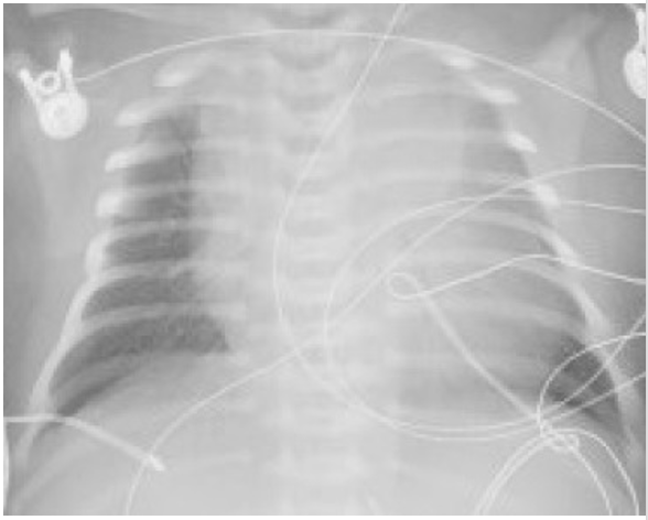
You order a blood gas test and a chest x-ray. The neonate’s x-ray is shown here. What is the pathophysiology of the most likely diagnosis?
A.Bronchospasm secondary to reactive airway disease
B.Vascular congestion of the epiglottis resulting in airway obstruction
C.Alveolar collapse related to increased forces of lung recoil
D.Pulmonary edema related to PDA
E.Hypoxia due to persistence of fetal circulation
INSTRUCTIONS
- Step 1: Answer the question, providing medical reasoning to back up your answer. Your answer should be at least 3-5 sentences. DO NOT REFRESH YOUR BROWSER UNTIL YOU HAVE FINISHED YOUR ANSWER.
- Step 2: Now, refresh your browser, and respond to as many others in your team as you can. Responses should provide evidence of your thinking processes (not just "I agree", or "answer is X"). Regularly refresh your browser as others' responses come in.
- Step 3: Based on the discussion recorded in CGScholar, your team reporter should now enter your team's answer in Benware. The whole-class discussion of this application exercise will take place verbally as usual.


It's so hard to distinguish pulmonary edema from atelectasis
Reading x-rays is difficult, but it looks like possible diffuse congestion on the left side? Which would line up with D, but I'm not sure
i think it is C
since baby had chest retractions, that was probs compensation of chest wall trying to counteract collapsing lung
Could the left lung maybe have alveolar collapse because it's mainly just gray and not a lot of air seems to be getting in?
I am lacking in the ability to read x-rays, but it appears to me that the right lung is not filling the chest cavity. I chose C because it is the only one that truly indicates this x-ray findings. It does not appear that there is any edema because there would be more white due to fluid. It looks like the main problem is in one lung, so it probably would not be a problem with the epiglottis. I do not believe it would be related to A or E either
Based on the history from the last question I think C is probably the best answer as well.
Is that an abnormally large cardiac silhouette or is that more like pulmonary congestion in the left lung?
I don't know how to read X-rays very well, but it does seem the right side of the heart seems larger than normal, which makes me think of some sort of fluid overload, whether it being pulmonary edema or interstitial edema (D or C).
@Morgan hahaha you're so right I just realized I said heart
I don't think that is the heart as it appears too large in comparison to the chest cavity. I think it is the lung?
I was kind of thinking the same thing!